Using a Particle Accelerator to See Inside a 1,900-Year-Old Mummy
We’ve come a long way from 19th-century mummy “unwrapping parties.”

In 1834, renowned surgeon Thomas Pettigrew stood before a sold-out audience at the Royal College of Surgeons in London. The crowd agog, he leaned over an authentic Egyptian mummy, and gradually began to peel off the wrapping. As Egyptomania swept across the continent, these “unwrapping parties” had grown increasingly common, and people gathered in numbers to see preserved flowers plucked from beneath millennia-old armpits. The events were incredibly popular, but had the downside of being incredibly destructive to their precious centerpieces.
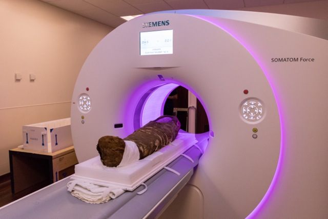
Today, modern medical imaging allows for “digital unwrapping” of mummies without damaging the contents within. Now, a team at Northwestern University has, for the first time, used high-energy synchrotron X-rays, generated by a particle accelerator, to look underneath the linen shroud of a 1,900-year-old child mummy.
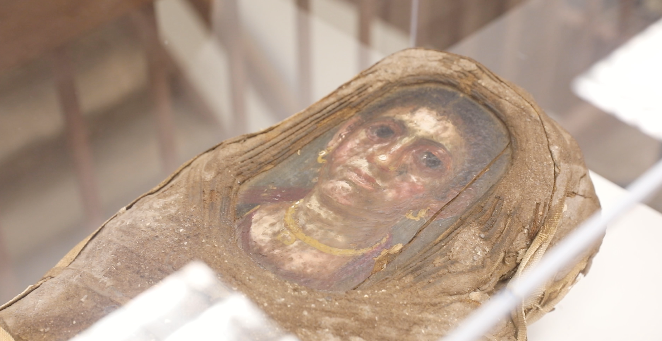
Found in 1911, the mummy contains the remains of a girl around five years old. Experts don’t know precisely who she was, but she received a lavish burial typical of the style of Roman-era Egypt. She is a rare “portrait mummy”—only 100 are known to exist—featuring a detailed, flat portrait positioned over the skull. These paintings are some of the oldest naturalistic likenesses in existence. The child’s portrait shows her gazing out, serene and melancholy. She wears a crimson tunic, and is adorned with gold jewelry. Her body, just three-feet-long, is swaddled in layer upon layer of linen arranged in a complex geometric pattern.
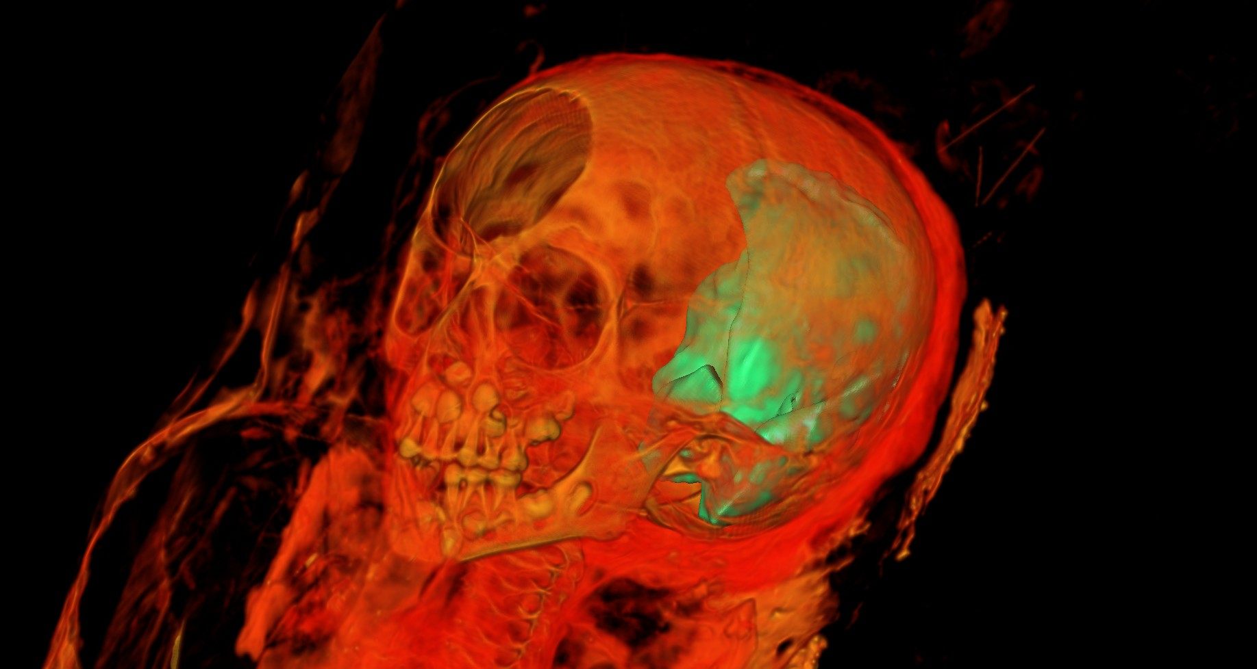
For this study, scientists at Northwestern employed a number of medical imaging technologies. The images reveal how the embalming process left a large hole and shards of what scientists say looks like solidified pitch in the brain cavity. Wires are wrapped around her head and feet. Her undescended adult incisors crowd for space above her milk teeth. Her arms are by her sides, her feet lightly folded over one another.
While there’s much more work to be done with the massive quantity of data the scans have produced, they should provide more clues about the girl and the time she lived in, said Northwestern research professor Marc Walton, in a statement. “This is a once-in-a-lifetime opportunity for our undergraduate students—and for me—to work at understanding the whole object that is this girl mummy,” he added. “Today’s powerful analytical tools allow us to nondestructively do the archaeology scientists couldn’t do 100 years ago.” The results are every bit as dramatic and wondrous as they were in the 19th century.


Follow us on Twitter to get the latest on the world's hidden wonders.
Like us on Facebook to get the latest on the world's hidden wonders.
Follow us on Twitter Like us on Facebook